The Clinical Relevance of Broad Mutational Screening of Myeloproliferative Neoplasms at Diagnosis

August 11, 2023

Helna Pettersson, Jenni Adamsson, Peter Johansson, Staffan Nilsson, Lars Palmqvist, Bjorn Andreasson, Julia Asp

Introduction: Myeloproliferative neoplasm (MPN) is a heterogenous group of hematological malignancies including polycythemia vera (PV), essential thrombocythemia (ET) and primary myelofibrosis (PMF). JAK2V617F is the most frequent driver mutation in all three entities, but in PMF and ET mutations in CALR and MPL are also frequent. Mutations seen in additional genes are also often the same regardless of subtype of MPN. The aim of this study was to analyze a population based MPN cohort for genetic variants with prognostic value that can guide clinical decisions.

Methods: MPN patients from Western Sweden diagnosed between 2008-2013 (n=248) were screened for mutations in 54 genes associated with myeloid malignancy.

Results: Mutations in the genes SRSF2 and U2AF1 correlated significantly with impaired overall survival but did not correlate to increased risk for vascular events, neither before nor after diagnosis. Rather, mutations in these genes showed an association with disease transformation. Several recurrent gene variants with allele frequency close to 50% were confirmed to be germline. However, none of these variants was found to have an earlier onset of MPN.

Discussion: In conclusion, we identified gene mutations to be independent markers of impaired survival in MPN. This indicates the need for more individualized assessment and treatment of MPN patients and a wider gene mutation screening already at diagnosis. This could ensure the identification of patients with high-risk mutations early on. In addition, several genetic variants were also identified as germline in this study but gave no obvious clinical relevance. To avoid conclusions from non-informative genetic variants, a simultaneous analysis of normal cell DNA from patients at diagnosis should be considered.

Introduction

Polycythemia Vera (PV), Essential Thrombocythemia (ET) and Primary Myelofibrosis (PMF) all belong to the Philadelphia chromosome negative myeloproliferative neoplasm (MPN) category. These three entities share the same characteristics of causing proliferation of bone marrow cells, resulting in an increase of blood cells of myeloid lineage in the bone marrow and in peripheral blood. Advanced stages of PMF, on the other hand, is characterized by increase of reticulin fibers leading to decreased blood cells (13). The complications of these three entities are also similar regarding vascular events, i.e., thrombosis and bleeding. Furthermore, all three entities can transform into acute leukemia and have an impact on survival, however with large differences in frequency. Despite the common driver mutations in JAK2, CALR and MPL (4), the clinical presentation, risk and frequencies of complications and survival differ wildly between individual patients. Prognostic tools are therefore desired in clinical practice for follow-up and treatment decisions already at diagnosis. Modern sequencing techniques have given the opportunity to simultaneously analyze several mutations in blood malignancies. It has become widely used in both research and clinical practice (5). We and other groups have published data on risk mutations using this approach, but several studies analyze data on separate MPN entities or driver mutation groups. Since both driver mutations and several of the additional mutations found are shared between the subtypes of MPN, and since the occurrence of some additional mutations are rather rare, we hypothesize that analysis of mutations in MPN as one group has a potential to extend the prognostic value of genetic markers. Furthermore, there is a growing number of hereditary gene variants that have been linked to predisposition for development of hematological disorders, including MPN (611). This also represents a challenge when analyzing large amount of sequencing data, especially if comparison with normal non-malignant cells is not available. We also need to get more information regarding gene variants of unknown significance, to avoid overestimation of their importance but also to identify variants that might influence disease development and prognosis. In this study, we analyzed a well-defined, population based MPN cohort, regardless of subtype, for genetic variants. The aim was to search for additional prognostic markers that can be used to guide clinical decisions, as well as to investigate the potential impact of germline variants detected in the sequence data.

Materials and methods

Patients

All patients diagnosed with PV, ET or PMF according to the 2008 WHO diagnostic criteria (3) in Western Sweden at the Sahlgrenska University Hospital or NU Hospital Group between 2008 and 2013 and reported to the Swedish national blood cancer registry were identified. Basically, all patients in our health care region with suspected MPN are referred to and treated at these two hospitals. Thus, this cohort cover patients in the geographic area without any selection. Of this cohort, 248 patients were included in the study based on informed consent, availability of DNA sample from the time of diagnosis, and review of diagnosis. Data from a subset of this patient cohort have been published previously (1213). Details are outlined in Supplementary Table 1. Clinical characteristics, clinical course, vitality status, vascular complications, disease transformation and co-existing cancers were collected from the medical records of all patients. Each of the patient’s hospital records were searched after emergency care consultation and hospital admission records that is related to bleeding or thrombotic complications. Follow-up was done from diagnosis until June 2021. The study was performed in accordance with the Declaration of Helsinki after ethical approval.

Screening for myeloid mutations

Genomic DNA from whole blood from the same sample that was analyzed at diagnosis for the presence of JAK2CALR or MPL mutation was screened for gene variants in 54 genes or mutational hot spots associated with myeloid malignancies. The TruSight Myeloid Sequencing panel (Illumina FC-130-1010) which also was used for diagnostics in the clinical laboratory at the time of the study, was used, and sequencing was performed on a MiSeq instrument (Illumina) according to manufacturer’s instructions. Secondary analysis was performed with MiSeq Reporter, v.2.4.60.8, using Burrows-Wheeler Aligner mapper and somatic variant caller (Illumina). Data were filtered and mapped to the human genome reference hg19 using Variant studio v3.0 (Illumina), where global filtering was set to >3% and coverage had a minimum of 500 reads. Variants causing missense, frameshift, an altered stop/initiation codon, in-frame insertion/deletion or variants affecting splice site were regarded as mutations. Variants with quality >Q30 and allele frequencies of at least 5% were considered positive for mutation. Known sequencing artefacts and variants previously found in normal controls were excluded from further analysis according to filter strategies used in the clinical laboratory. BAM files from secondary analysis were used to analyze selected variants by Integrative Genomics Viewer (www.broadinstitute.org). Variants in areas with difficult reads were excluded. Previously analyzed data was reanalyzed according to updated bioinformatic settings to make the results comparable regardless of time for sequencing.

Confirmation of germline variants

Blood sample was taken from patients with variants in CDKN2A (rs3731249), ETV6 (rs145477191), NOTCH1 (rs61751489) or MPL (rs41269541), with a variant allele frequency close to 50%. Blood was enriched for CD3+ cells, using MACS® cell separation kit StraightFrom™ Whole Blood CD3 MicroBeads (Miltenyi Biotech) and Whole Blood Column Kit (Miltenyi Biotech), according to the manufacturer’s protocol. Genomic DNA from CD3+ enriched cells were extracted using QIAamp DNA Blood Mini Kit (Qiagen) and 10 ng of DNA were genotyped using TaqMan SNP genotyping assay (Applied biosciences, Life technologies) according to manufacturer’s protocol. The following assays were used: CDKN2A (assay ID: C_25611114_10), ETV6 (assay ID: C_162058060_10), NOTCH1 (assay ID: C_90123839_10) and MPL (assay ID: ANFV4EK). All samples were analyzed in triplicates using the QuantStudio 3 Real-Time PCR system (ThermoFisher Scientific). Genotypes were determined automatically based on dye component fluorescent emission data depicted in the X–Y scatter plot using Taqman genotyper software v.1.6.0. The gnomAD database v2.1.1 (https://gnomad.broadinstitute.org/) was used to compare frequencies in the MPN cohort with a normal Swedish population.

Statistical analysis

Fisher’s Exact Test was used to compare differences in frequencies between groups. To estimate overall survival (OS), defined as time from diagnosis to last follow up or death from any cause, the Kaplan Meier Log-rank test was used initially. For multivariable analysis, logistic regression and Cox Regression was used. P-values <0.05 were considered statistically significant. The statistical software used were Analyze-it v.6.15.4 (Microsoft Excel), GraphPad Prism v.9.4.0 and SPSS v29.0.0.0.

Results

A population-based cohort

Between 2008 and 2013, 300 patients were diagnosed with MPN at Sahlgrenska University Hospital and NU Hospital Group in Western Sweden. Of these, 83% (n=248; PV n=84, ET n=123, PMF n=41) were included in the study. All included patients fulfilled the 2008 WHO diagnostic criteria. Age, gender, and blood counts from the time of diagnosis, for the whole MPN group and for the sub entities, are presented in Table 1. The distribution of driver mutations found at diagnosis was consistent with expected findings in the different subgroups of MPN (Figure 1). One patient with PMF was found to harbor both JAK2 V617F as well as mutation in the CALR gene. Patients not included in the study either declined to participate, had another diagnosis when their medical records were reviewed, or diagnostic material was missing. The median age of these patients was slightly lower (66 years vs. 69 years) but there was no other significant difference between these and the included patients.

Table 1
www.frontiersin.org
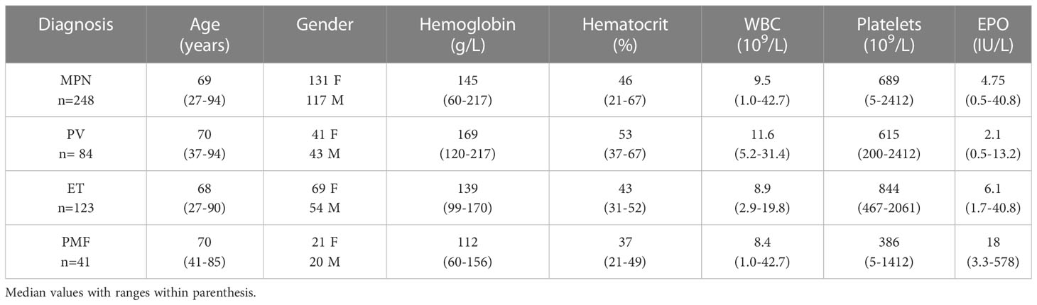
Table 1 Age, gender and laboratory findings at diagnosis in 248 patients with MPN.

Figure 1
www.frontiersin.org
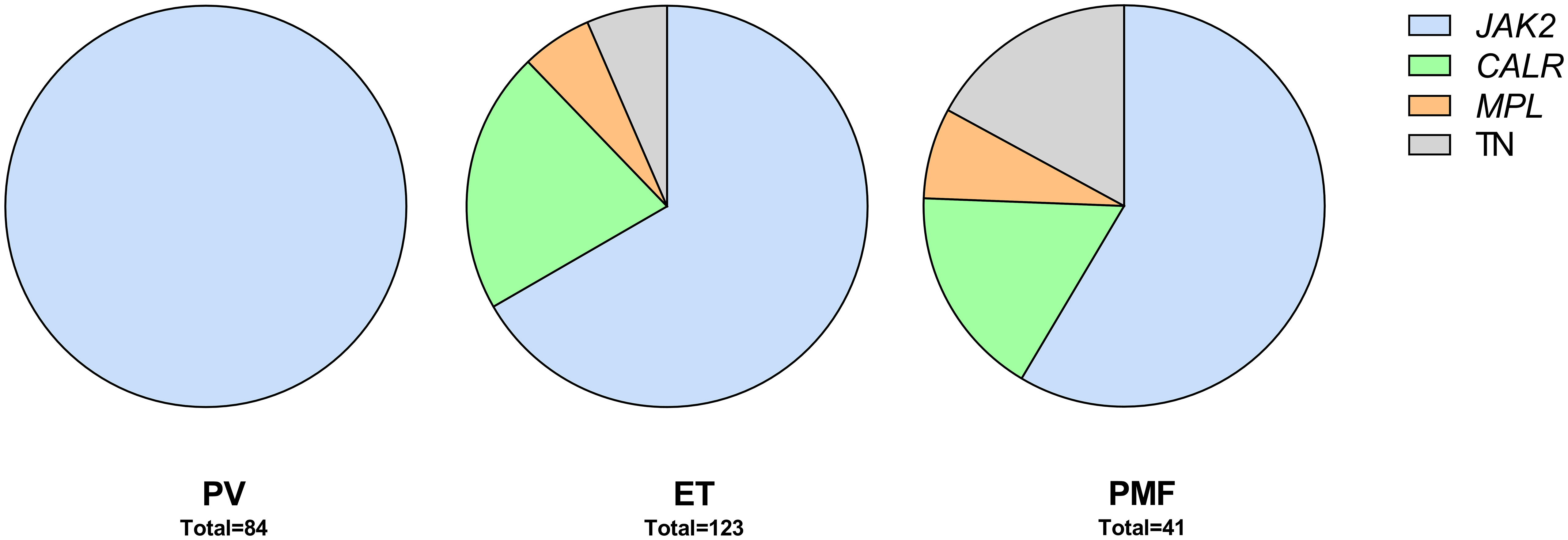
Figure 1 Patients by diagnosis and driver mutations. TN, triple negative.

Mutations and survival

A sequencing panel including 54 genes associated with myeloid malignancies was used to screen for genetic variants that could be used as prognostic markers. Variants regarded as mutations other than the diagnostic driver mutations (JAK2CALR or MPL) were found in 37 genes in at least one patient and in 27 genes in at least three patients (Figure 2ASupplementary Table 1). During analysis of the gene data, several recurrent gene variants with allele frequency close to 50% were noted, which implied a hereditary variant. Therefore, analysis of the most common variants CDKN2A (NM_001195132.1:c.442G>A), NOTCH1 (NM_017617.3:c.6853G>A) and ETV6 (NM_001987.4:c.602T>C) as well as a variant close to a splice site in the MPL gene (NM_005373.2:c.1565 + 5C>T) were also analyzed in separated T-cells from new blood samples. This confirmed the variants to be germline. Therefore, these variants were excluded from analyses of prognostic impact. Sixty-three percent of the MPN cases had other mutations in addition to the diagnostic driver mutation (Figure 2B). Presence of at least one additional mutation was found to be associated with inferior survival (Figure 2C). To investigate if it was mutations in general or mutations in particular genes that had impact on survival, all genes with mutations detected in at least three patients were correlated to survival with the Kaplan Meier Log-rank test. For the whole MPN group, only mutations in five genes correlated significantly with inferior overall survival, ASXL1 (P=0.0005), SRSF2 (P<0.0001), U2AF1 (P<0.0001), CBL (P=0.01) and SF3B1 (P<0.0001) (Figure 3). These were further tested with multivariable analysis using Cox regression where also age and type of diagnosis were taken into consideration. When the five genes were grouped together, they still correlated to OS (P=0.002) with a hazard ratio 3.248, and it was not dependent on type of diagnosis (interaction 0.592). Also, age at diagnosis correlated to OS (P<0.001) as expected. However, it should be noted that all cases with CBL mutation also harbored mutation in another of the four genes (Supplementary Table 1). Therefore, the genes were also tested separately. When these were adjusted for both age and type of diagnosis, only mutations in SRSF2 and U2AF1 correlated significantly to OS (Table 2).

Figure 2
www.frontiersin.org
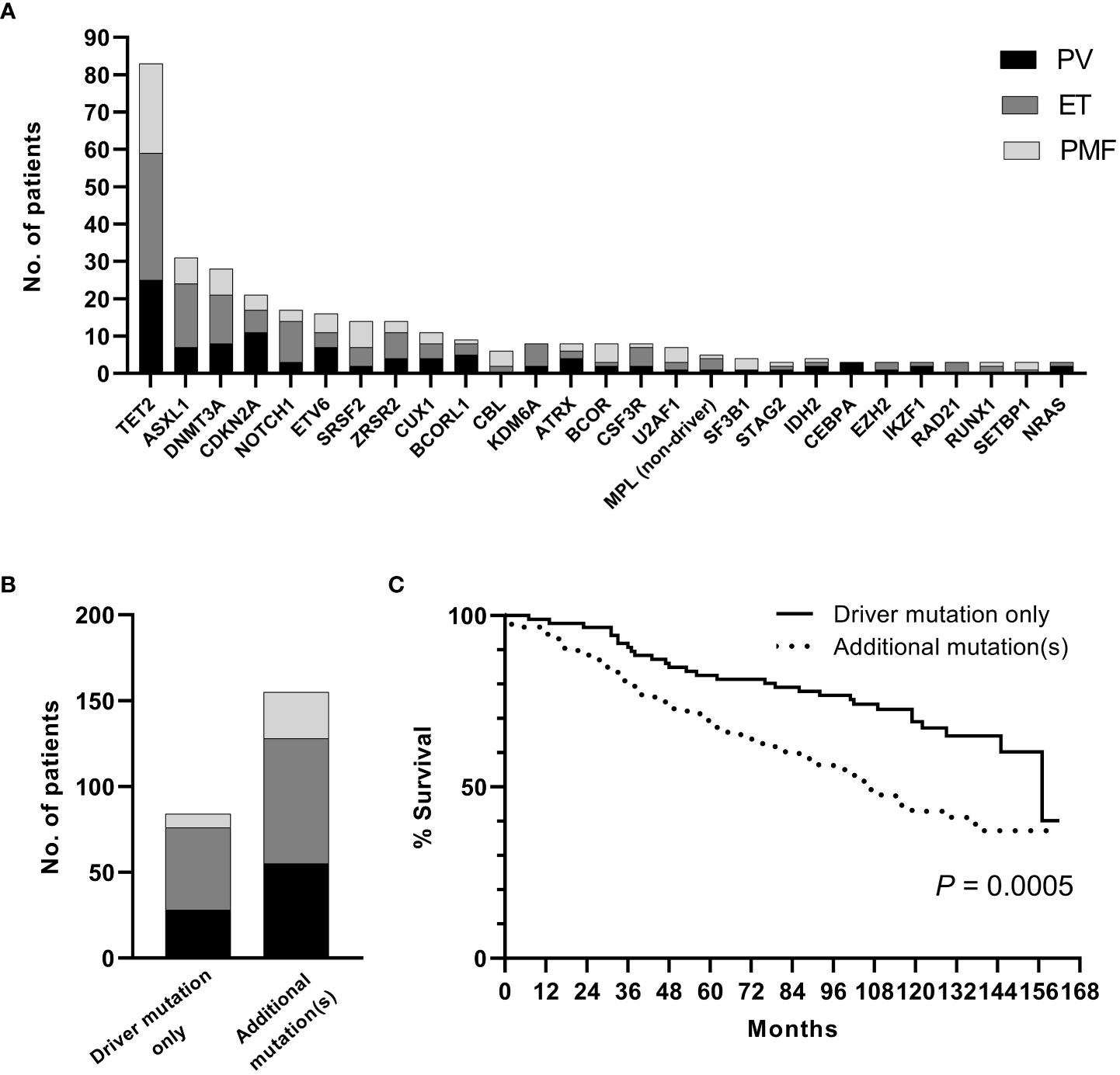
Figure 2 (A). Frequency of additional mutations. (B). Distribution of patients with driver mutation only or addition mutation(s) when confirmed germline variants have been excluded. (C). OS in patients with driver mutation only or with addition mutation(s) (confirmed germline variants excluded).

Figure 3
www.frontiersin.orgFigure 3 OS in patients with mutations in ASXL1CBLSF3B1SRSF2 and U2AF1 respectively according to Kaplan Meier Log-rank test.

Table 2
www.frontiersin.orgTable 2 Mutation impact on OS in univariate or adjusted (including age and type of diagnosis) analysis.

Mutations and vascular events

Vascular events in MPN are potentially life-threatening. The vascular complications are either thrombosis or bleeding where the co-existence of MPN is a contributing factor. The most common incidences are those that are discovered at the time of diagnosis and the most common thrombotic events were myocardial infarction (n=27), cerebrovascular infarction (n=24), pulmonary embolism (n=19), transient ischemic attack (n=10) and deep vein thrombosis (n=8). The most frequent hemorrhagic complications were gastro-intestinal (n=11) and cerebral bleedings (n=6). Fisher’s Exact Test and logistic regression were used to analyze if SRSF2 or U2AF1 which correlated with shorter OS also correlated with occurrence of vascular events before or after diagnosis or in total. However, no such correlation was seen, neither when only the mutated genes were tested, nor when they were combined with age and type of diagnosis.

Mutations and disease transformation

All MPNs have a risk of transformation into secondary acute myeloid leukemia (AML). In our cohort, 17 cases had transformed to AML. Both mutated genes with correlation to OS were tested with logistic regression. This showed that mutations in SRSF2 correlated with AML transformation (P=0.002), but this was not the case for U2AF1 (P=0.236). The analysis was extended to find genes correlated to fibrotic transformation and co-existence with other myeloid hematological malignancies. There were 18 patients that had secondary myelofibrosis transformation from PV and ET. Other myeloid hematological malignancies that co-existed with MPNs included two chronic myelomonocytic leukemia and one myelodysplastic syndrome. Logistic regression showed that mutations in both SRSF2 and U2AF1 correlated with co-existing myeloid hematological malignancies (SRSF2 P=0.05 and U2AF1 P=0.014).

Gene germline variants

Identified germ line variants indicated a possible hereditary predisposition of MPN. Comparison of the frequency in our MPN cohort to a normal Swedish population cohort in the gnomAD variant database was performed. Only the variant found in ETV6 was more frequent in the MPN group (0.0282 vs. 0.00975 in allele frequency). This difference was statistically significant (Fischer’s exact test, p=0.0006). However, there was no correlation between any of the variants and occurrence of early onset MPN. We further used logistic regression to test if any of the variants correlated with occurrence of other cancers (both solid tumors and hematological malignancies outside the MPN group). These cancers occurred about the time and after diagnosis of MPN and were noted upon reviewing the patient´s hospital records. In total, 19 patients with non-hematologic cancers were found. The most common types were colon cancer (n=6) and pancreatic cancer (n=4). However, no significant correlation was seen.

Discussion

The serious risks all MPNs impose, although at various frequencies, are vascular complications, transformation to more severe hematologic malignancies and ultimately negative impact on OS. It is thus a priority to identify high risk patients in clinical practice. Age at diagnosis as well as occurrence of vascular complications have been reported as risk factors (1415). Access to an abundance of genetic data allows genetic profiling to further broaden prognostic information. Mutational status has progressively taken a big role in clinical practice. Occurrence of mutations have also been used to create scoring systems for MPN (1617). The initial focus on gene mutations in MPN was on the driver mutations’ importance on disease development. These mutations are found in the genes JAK2CALR and MPL which all are involved in JAK-STAT signaling (18). Notably, the same JAK2 mutation is found in both PV, ET and PMF, and mutations in CALR and MPL are seen in both ET and PMF. Thus, the mutation itself does not seem to determine the MPN phenotype, instead, allele burden has been reported as one factor behind the phenotypic differences (19). The order of acquisition of the driver mutation in relation to additional mutations may also have influence (2021). If the JAK2 mutation precedes mutation in DNMT3A or TET2, the phenotypic picture would likely be PV. If mutations instead occur in reverse order, the MPN phenotype would likely be ET (22). Host factors also contribute to the development of disease (2223). Several predisposing gene variants have been identified that may influence not only the risk of developing disease but also the course of the disease (172426). It is therefore reasonable to investigate the whole MPN cohort as a group independent of diagnosis when analyzing it from a genetic point of view.

Aside from the driver mutations, several additional mutations have also been reported in MPN. These are subclassified to gene families: epigenetic regulators (ASXL1, EZH2, TET2, IDH1/2, DNMT3A), spliceosome (SRSF2, SF3B1, U2AF1, ZRSR2), transcriptional regulators (TP53, RUNX1, IKZF1), general cell signaling genes (KRAS, PTPN11) as well as specific negative regulators of JAK/STAT signaling (CBL) (27). Occurrence of additional mutations correlated significantly with inferior OS (Figure 2C). Genetic profiling has raised the question if the number of mutations in a particular case is more important for the occurrence of complications or OS rather than in what genes or type of gene the mutations are present. Our results suggest it is not the number of mutations but rather the presence of certain gene mutations that are more informative for prognostic guidance. Mutations in SRSF2 and U2AF1 correlated significantly with worse OS in our patient cohort. Although they were more frequent in PMF, which is well known to have an impaired survival compared to patients with PV and ET, mutations in these two genes correlated to worse OS regardless of MPN subtype.

Previous studies, including a subpopulation of our analyzed MPN cohort, have shown that triple negative MPN without JAK2CALR or MPL mutation have worse prognosis (1228). It has also been shown that the presence of several other mutations in addition to a driver mutation correlate with survival (2524262832). In this study we initially identified mutations in five non-driver genes (ASXL1SRSF2U2AF1SF3B1 and CBL) to be significantly correlated to impaired OS. Mutations in both ASXL1 and SRSF2 have previously been classified as high-risk mutations in both PMF and PV (2633). Moreover, mutations in U2AF1 and SF3B1 have been identified as genetic risk factors in ET (17). When age at diagnosis as well as type of diagnosis was taken into consideration only mutations in SRSF2 and U2AF1 remained associated with shorter OS. Mutations in CBL were only found in those patients who harbored mutations in one or more of the four other genes, suggesting that mutated CBL might just be a passenger rather than a disease driver. Mutations in ASXL1 is commonly seen in clonal hematopoiesis, which increases with age (34). This could be an explanation why the presence of ASXL1 mutation no longer significantly correlated with OS when age was taken into consideration.

Since vascular complications are associated with impaired survival we wanted to investigate if the detected mutations correlated also to vascular events in our MPN cohort. However, neither mutations in SRSF2 nor U2AF1 correlated to vascular events before or after diagnosis or in total. Another complication with MPN is transformation to myelofibrosis for PV and ET or to secondary AML for all three MPN. In the present cohort, a significant correlation between mutations in SRSF2 and transformation to AML was found. Furthermore, mutations in both SRSF2 and U2AF1 correlated with transformation from PV and ET to myelofibrosis and development of other hematological malignancies. This is in line with previous findings were mutations in SRSF2 and U2AF1 have been reported to serve as prognostic markers for rapid blastic progression in newly diagnosed MPN (35). Moreover, mutations in SRSF2U2AF1 and SF3B1 detected at presentation of disease have been associated with rapid fibrotic progression in PMF. This was not demonstrated for mutations in ASXL1DNMT3A or TET2 (36).

Aside from the acquired mutations in our MPN cohort, several specific variants were identified which turned out to be germline. A five- to sevenfold higher risk of MPN among first-degree relatives to MPN patients have previously been reported in Sweden, which suggest a genetic predisposition (37). Also in other myeloid malignancies, the question for germline variants involved in disease have come into focus (3840). Four variants in our study were more closely investigated, their allele frequency was close to 50%, which could imply a hereditary variant. These genes were CDKN2A (NM_001195132.1:c.442G>A), NOTCH1 (NM_017617.3:c.6853G>A) and ETV6 (NM_001987.4:c.602T>C) as well as a variant close to a splice site in the MPL gene (NM_005373.2:c.1565 + 5C>T). The most frequent occurring CDKN2A mutation leading to a p.A148T substitution has been reported as an inherited coding variant associated with leukemic transformation of hematopoietic progenitor cells (41). Comparison of the frequency to a normal Swedish population cohort, however, only revealed the ETV6 variant to be more common in the MPN patient cohort. This variant did not correlate to earlier onset of disease, which could be expected for an inherited predisposition. On the other hand, in the Landgren study the mean age at diagnosis did not differ between affected relatives and controls (37). The ethical approval of the current study did not include testing of relatives, but it would of course be of interest to see if any of these variants are associated with an increased incidence of hematological or non-hematological malignancies within these families. Nevertheless, it is important to correctly identify germline gene variants to avoid drawing conclusions from non-informative genetic variants but also to provide genetic counseling when called for. In this study we used CD3+ selection of T-cells from collected blood samples to get constitutive DNA, which turned out to be easiest for both referring doctors and gave acceptable DNA yield for the laboratory but might of course misdiagnose somatic variants that are also present in lymphoid cells. Another alternative is a skin biopsy but this may be considered too much of an intervention for some patients.

In conclusion, our study on a population based MPN cohort strengthens previous reports about prognostic value of genetic data in MPN. Thus, a wider gene profiling at diagnosis is of value. In addition, several genetic variants were also identified as germline in this study but gave no obvious clinical relevance. To avoid conclusions from non-informative genetic variants, simultaneous analysis of normal cell DNA from patients at diagnosis should be considered.

Read more

What Is Myelofibrosis?

By Julie Scott, MSN, ANP-BC, AOCNP

Published on July 26, 2023

Myelofibrosis is a rare type of bone marrow cancer. In this condition, extensive scarring (fibrosis) occurs in the bone marrow, which keeps the bone marrow from producing the right number of blood cells.1

When myelofibrosis occurs, some people may not have any symptoms, while others have severe symptoms that require immediate treatment. This article will explain the symptoms of myelofibrosis, how it is diagnosed, and how it is treated.

How Myelofibrosis Affects the Body

Bone marrow is present inside the middle of the bones. It is normally a soft, spongy texture. It produces WBCs, RBCs, and platelets.

In myelofibrosis, one of the cells of the bone marrow begins to grow abnormally, multiply, and continue to produce more abnormal cells. Eventually, abnormal cells are present in high enough numbers to crowd out the healthy cells.

These abnormal cells cause fibrosis, which prevents the bone marrow from producing the correct number of blood cells the body needs to function normally.2 Over time, there is an increased risk of developing acute myeloid leukemia (AML), another form of blood cancer.1

Myelofibrosis Types

The initial cause of abnormal bone marrow development determines the type of myelofibrosis. The two types are primary and secondary.

Primary Myelofibrosis

With primary myelofibrosis, the change in the bone marrow cells happens spontaneously. It doesn’t occur due to a previous bone marrow condition.1

Secondary Myelofibrosis

With secondary myelofibrosis, the fibrosis occurs due to another bone marrow disorder, specifically polycythemia vera or essential thrombocythemia.3

Polycythemia vera is a blood disorder in which the bone marrow produces too many blood cells, most often red blood cells, but can also include white blood cells and platelets.2 Essential thrombocythemia is a disorder in which the bone marrow makes too many platelets.4

Myelofibrosis Symptoms

As too few blood cells are made, symptoms will start to develop. The rate at which symptoms develop and how severe they become can vary from person to person, and may take years to be experienced. Symptoms associated with myelofibrosis include:2

  • Feeling tired
  • Shortness of breath
  • Pale skin
  • Headaches
  • Fever
  • Night sweats
  • Enlarged spleen
  • Enlarged liver
  • Frequent infections
  • Easy bleeding or bruising
  • Abdominal pain
  • Joint pain
  • Bone pain

Tumors may develop in the lungs, skin, liver, or gastrointestinal tract and cause further symptoms.2

Causes of Myelofibrosis

For those living with primary myelofibrosis, the exact cause of the disease may never be known.

However, in about half of the cases of primary myelofibrosis, a mutation in the JAK2 gene is found.5 The JAK2 mutation is also frequently found in those with polycythemia vera and essential thrombocythemia.

This mutation isn’t inherited. Instead, it develops spontaneously in a bone marrow cell. It produces a protein that causes the bone marrow to overproduce platelet precursor cells called megakaryocytes. These cells stimulate other cells to produce too much collagen, a protein that then builds up and produces scarring in the bone marrow.

Other gene mutations that may play a role in developing myelofibrosis include the CALR and MPL genes.1

Risk factors that may play a role in developing primary myelofibrosis include:2

  • Increasing age
  • History of exposure to chemicals including benzene, fluoride, or phosphorus

Risk factors for developing secondary myelofibrosis include:6

  • Having another cancer that has spread into the bone marrow
  • Having polycythemia vera or essential thrombocythemia

Diagnosis of Myelofibrosis

The diagnosis of myelofibrosis often starts when someone presents to their healthcare provider for evaluation of a symptom that they are experiencing. During this evaluation, a healthcare provider may start with a detailed history and physical examination. Blood work may be taken which can start the process of finding a diagnosis.1

A complete blood count (CBC) and peripheral blood smear measure the number of WBCs, RBCs, and platelets, as well as their shape and size. Abnormal findings in the CBC may lead to further testing, which may include a bone marrow biopsy.1

During a bone marrow biopsy, a small sample of bone marrow is taken, often from the hip. This allows the pathologist (physician specializing in analyzing body fluids and tissues in a lab setting) the ability to evaluate for any changes or abnormalities in the bone marrow. This test can result in a diagnosis of myelofibrosis.

In addition, a physical exam or imaging study such as a computed tomography (CT) scan may reveal an enlarged spleen.

Other blood testing may include:1

  • Complete metabolic panel (CMP) to evaluate kidney and liver function
  • Coagulation studies
  • Iron levels
  • Lactate dehydrogenase (LDH) to assess inflammation and tissue damage
  • Testing for the JAK2, CALR, and MPL gene mutations

Once a diagnosis of myelofibrosis is made, it is further classified into different risk categories, which helps determine how likely the disease is to turn into AML and can help determine treatment options.

This score is determined by the person’s age, symptoms, hemoglobin level, platelet count, leukocyte (a white blood cell) and leukoblast (a developing white blood cell) count, and certain genetic changes. The higher the score, the more high-risk their myelofibrosis is.

Myelofibrosis Treatment

Some people diagnosed with myelofibrosis, especially those without many symptoms or who have low-risk disease, may not receive any treatment until they become symptomatic. Called a watchful waiting approach, this policy of taking no immediate action regarding treatment includes routine blood tests and visits with their healthcare provider to determine when treatment will be needed.2

If someone is experiencing symptomatic anemia (low RBCs) because of myelofibrosis, they may receive periodic RBC transfusions. There are additional medications that may be given to help the bone marrow make red blood cells.

This may not completely resolve anemia but can keep the red blood cells up at a tolerable level. If someone also has iron deficiency anemia, iron supplements may help improve red blood cell levels.2

To reduce high levels of WBCs and platelets, medications to suppress the bone marrow may be given. An example of one of these medications is hydroxyurea.

An enlarged spleen may need to be treated if it is contributing to symptoms, especially pain or severely low platelets. This can be done through radiation to the spleen or by surgical removal of the spleen.

A medication called Jakafi (ruxolitinib) can be prescribed to those with either primary or secondary myelofibrosis who fall in the moderate- or high-risk category. This medication interferes with the JAK2 pathway that the cells use to grow. Another medication, Inrebic (fedratinib) can also be used to treat intermediate or high-risk primary or secondary myelofibrosis.2

Can Myelofibrosis Be Cured?

The majority of cases of myelofibrosis are treated with the goal of decreasing symptoms of the disease. An attempt can be made to cure the disease through a stem cell transplant.

This approach requires large doses of chemotherapy to kill all of the cancer cells. Stem cells are collected before the procedure to be infused back in after chemotherapy has worked. These stem cells can then begin to resume making normal WBCs, RBCs, and platelets. This procedure is not recommended for everyone with myelofibrosis, as it can lead to severe complications.7

Complications Associated With Myelofibrosis

Complications associated with myelofibrosis are related to the severe decrease in the number of normal WBCs, RBCs, and platelets. As the disease progresses and the blood counts continue to decrease, complications may arise.

With the decrease of white blood cells comes a higher risk of developing infection. Infections can occur anywhere in the body, though most often in the lungs. The infection can be due to bacteria, viruses, or fungi. With infection may come fever, increased weakness, and cough.8

Low red blood cells can result in severe anemia, which prevents enough oxygen-rich blood from getting to the tissues in the body. With the decreased amount of available oxygen comes complications such as heart failure, in which the heart has to work too hard to try to keep up with the increased demand for oxygen.8

Not having enough normal platelets can lead to severe bleeding or hemorrhaging. The bleeding can occur following an injury or can occur spontaneously. The bleeding can become life-threatening if severe and not stopped quickly.8

Blood clotting, the opposite of bleeding, could also occur. If blood clots inappropriately, it can lead to clots moving around the body and getting stuck in areas they are not supposed to be. This can lead to damage to the brain, heart, lungs, and extremities.

Transformation to acute myeloid leukemia occurs in 5% to 10% of those diagnosed with myelofibrosis. This is most common in primary myelofibrosis and is a significant complication since the prognosis is poor with transformation into AML.9

Myelofibrosis Prognosis

The prognosis of myelofibrosis can vary from person to person. It depends upon the type and risk category of their disease. Prognosis can differ slightly based on which scale is used at the time of diagnosis. The table below references the prognosis scale MIPSS70, which is used for those 70 years old or younger and is based on risk group severity.9

 Risk Group 10-Year Survival
 Very high Less than 5%
 High  13%
 Intermediate  37%
 Low  56%
Very Low  92%

When to Contact a Healthcare Provider

See a healthcare provider if you are having symptoms associated with myelofibrosis. Many of these are also associated with other conditions. A workup and diagnosis can ensure you are getting the appropriate treatment.

If you have been diagnosed with myelofibrosis, contact your healthcare provider anytime you’re having concerns about the symptoms you’re experiencing, especially if they continue for some time without getting better.

Your provider may want to run additional tests or start treatment if your symptoms continue. If severe symptoms develop, notify your healthcare provider immediately or seek emergency care.

Summary

Myelofibrosis is a type of blood cancer in which abnormal cells cause the bone marrow to become extensively scarred (fibrosis). The fibrosis doesn’t allow the bone marrow to make blood cells properly, which leads to low blood counts and other complications.

Once formally diagnosed by a bone marrow biopsy, the results will be used by your healthcare provider to develop a treatment plan. Treatments are individualized, ranging from watchful waiting to stem cell transplant.

Read more

Know What Questions to Ask When Treating MPNs

July 27, 2023

Brielle Benyon

Oncology nurses must know what to look for — and what questions to ask — when treating patients with myeloproliferative neoplasms (MPNs), as side effects and other patient characteristics can play a role in determining the best treatment regimen, according to Julie Huynh-Lu, PA-C, a physician assistant from The University of Texas MD Anderson Cancer Center.

MPN-Related Symptoms

“Whenever a patient comes to see us, they also fill out the MPN10 questionnaire, [which is] a list of all the 10 symptoms that frequently occur in our patients,” Huynh-Lu said in an interview with Oncology Nursing News. “Ideally, this should be occurring at every visit. On top of them filling that out, I obviously will ask them specific pointed questions as well just to tease out some more information. But this should occur at every visit.”

Symptoms can vary based on the subtype of MPN a patient has. Huynh-Lu said that patients with polycythemia vera and essential thrombocytosis are more likely to experience headaches, confusion or difficulty focusing, or pain and tingling in the fingertips. Meanwhile, common symptoms for patients with myelofibrosis include anemia and thrombocytopenia; shortness of breath and fatigue; bleeding; and complications from spleen enlargement, such as having a poor appetite.

Knowing about these symptoms is key, as they could indicate a physical issue that warrants a change in treatment, Huynh-Lu said. For example, if a patient is not experiencing splenomegaly (enlarged spleen), there may not need to be prescribed a JAK inhibitor. However, if the patient starts to experience a decreased appetite or feel full after eating only a small amount of food, that could indicate that their spleen is becoming enlarged, and that patient may benefit from being put on a JAK inhibitor.

“It can also change the trajectory on whether or not talking about splenectomy is an option. It’s not really our go-to [treatment] in our department at MD Anderson, but that could certainly lend to a conversation into if surgery is an option,” Huynh-Lu said.

Sometimes symptoms can lead to a change in treatment, while other times there may be an easy fix to manage the issue.

If a patient is currently taking a JAK inhibitor, nurses should be sure to ask them about worsening itching, diarrhea, and frequent infections (such as urinary tract infections or pneumonia). Secondary skin cancers can also occur, said Huynh-Lu, “so we always recommend that they get dermatology checks every 6 months.”

“If their [blood] counts are starting to drop, or if their spleen is starting to grow, well, maybe the medication they’re on right now, the dosage needs to be altered. But if we alter the dose to a higher medication dose, and the side effects are worse, maybe then it’s time to switch to a different class of drugs completely, or same class of drugs, just a different type of drug. There’s also, of course, clinical trials that are available, so that could be an option as well,” Huynh-Lu said.

Patient Characteristics and Comorbidities

Regarding patient characteristics and comorbidities, clinicians should know if patients have a history of cardiac, renal, or hepatic complications, as certain medications can affect these organs.

Additionally, interferons are commonly used to treat patients with polycythemia vera. However, according to the National Institutes of Health, these drugs can impact the synthesis of serotonin, dopamine, epinephrine, and norepinephrine, thereby increasing a patient’s risk for depression. That said, clinicians should know if patients have a history of depression or an autoimmune disease before they prescribe an interferon to a patient, Huynh-Lu said.

It also may be beneficial for oncology nurses to ask patients if they are experiencing financial struggles due to their cancer care.

“I know these drugs can be quite expensive. Financially, this can be a burden for some … Sometimes the local oncologists aren’t completely aware of financial assistance available for them. So maybe just ask and say, ‘Hey, I know this drug cost this much. Do you know of any financial assistance that you guys can provide for me?’ Because I know sometimes that’s not a question that gets asked,” Huynh-Lu said.

Read more

Patients and Providers Need to Stay Vigilant in Assessing, Managing MPN Symptoms

July 25, 2023

Alex Biese

Nurses play a crucial role in managing symptoms experienced by patients with myeloproliferative neoplasms (MPNs) — a group of blood cancers that cause the bone marrow to overproduce red or white blood cells or platelets.

“MPNs are rare, and the nurse/nurse practitioner [must] take the time to listen to the patients and educate them about symptom management,” Alfa Lafleur, APRN, from Florida Cancer Specialists and Research Institute Trinity Cancer Center in Trinity, Florida, told Oncology Nursing News®.

Even though the MPN category includes a range of diseases such as myelofibrosis, essential thrombocythemia, and polycythemia vera, patients with MPNs experience several common symptoms. Lafleur said she likes to think of those symptoms in categories.

First, Lafleur explained, there are the inflammation-related symptoms including weight loss, night sweats, fever, fatigue and a general feeling of unwellness. Then, there are the microvascular-related symptoms related to dysregulation of the JAK2 pathway, which can cause numbness in the hands and feet as well as headaches, vision changes and a painful rash. There are also symptoms related to the enlargement of the patient’s spleen, including fullness, pain and discomfort in the upper left quadrant of the abdomen.

As MPN symptoms typically intensify over time, with patients potentially experiencing long asymptomatic periods, the symptom identification and management work of a nurse is of particular importance.

“The nurse remains vigilant in assessing for symptoms as early identification that the disease may be worsening or transforming (which) can result in improved patient outcomes,” Lafleur said.

It’s also important for patients to keep tabs on their symptoms, as the MPN Research Foundation explains on its website.

“Because symptoms can vary significantly among MPN patients, it is important to track changes in symptoms and their severity between doctor visits,” the foundation states. “Keep current on the latest MPN research updates and speak to your doctor about how changes in your symptoms may call for a change in treatment.”

Resources are available for both patients and providers. Lafleur cited the MPN Research Foundation’s education for providers and patient support hotlines staffed by counselors who can in turn provide emotional support and assistance in locating additional resources and support groups.

Additionally, Voices of MPN, she said, “has a phenomenal app that helps the patient to track their symptoms, educates on the disease process and offers other resources and support to the patient.”

Patients with MPNs, Lafleur noted, can live with their cancer for years as they undergo different treatments that are each associated with side effects and symptoms — an experience that, she said, “can be really frustrating and upsetting” — and can be accompanied by fear of MPNs’ potential transformation into more aggressive forms of cancer such as leukemia or the risk of experiencing amyocardial infarction or cerebral vascular accident, or CVA, also known as a stroke.

Lafleur has some straightforward guidance for patients: “My best advice to patients is to remind them that they are not alone in this disease,” she said. “No symptom is too small to bring to the attention of your nurse who is more than willing to assist with the physical and mental challenges that come.”

Reference

MPN Research Foundation. Accessed July 25, 2023. https://www.mpnresearchfoundation.org/

Expert Panel Discusses Advocacy, Education for Patients With Myeloproliferative Neoplasms

Jul 14, 2023

Erin Hunter, Assistant Editor

Patients with hematologic myeloproliferative neoplasms (MPNs)—a group of rare blood diseases that include myelofibrosis, essential thrombocythemia (ET), and polycythemia vera (PV)—should take a more active role in their treatment plan, according to experts in oncology pharmacy who participated in a Pharmacy Times clinical forum at ASCO 2023 in Chicago, Illinois.

“I think that our role as pharmacists is to give [patients] as much information as we possibly can, and then encourage them to move forward with advocating for themselves,” said Krystal Preston, PharmD, BCPS, senior clinical specialist-oncology pharmacist, CVS Health, and professor, University of Chicago College of Pharmacy, Chicago, Illinois.

Patients who are serious about taking on an active role in their treatment could inspire health care providers to collaborate more both with them and other members of the care team, according to discussion leader Zahra Mahmoudjafari, PharmD, MBA, BCOP, FHOPA, clinical pharmacy manager of hematology, bone marrow therapy, and cellular therapeutics at the University of Kansas Health System in Mission, Kansas.

Myelofibrosis, ET, and PV are subtypes of MPNs. ET and PV typically transform into myelofibrosis, which can subsequently turn into acute myeloid leukemia (AML). At least 20% of MPNs may transform into AML, therefore, the goal for treatment is to prevent this from occurring, Mahmoudjafari explained.

More than 90% of patients with PV have a Janus kinase 2 gene (JAK2) mutation, “and it is probably, by far, the mutation that we have the most actionable ability to do something about,” Mahmoudjafari said. She added that because there are currently 3 FDA-approved JAK inhibitors for MPN—ruxolitinib (Opzelura; Incyte), fedratinib (Inrebic; Bristol Myers Squibb), and pacritinib (Vonjo; CTI Biopharma Corp.), which were approved based on results from the COMFORT-1, JAKARTA, and PERSIST-2 pivotal trials, respectively.

Ruxolitinib and fedratinib are primarily for patients with intermediate- or high-risk myelofibrosis, including intermediate-2, and primary and post-PV/ET myelofibrosis. Pacritinib is indicated for patients with a platelet count below 50,000, and all have an expected adverse events profile, which includes thrombocytopenia, anemia, bruising, dizziness, headache, and diarrhea.

The only true cure for myelofibrosis is transplant, however, there is a 30% mortality risk associated with it, Mahmoudjafari said. During the forum, panelists largely explored patient management. Compliance was cited as a predominant issue for Connor Roth, PharmD, BCOP, hematology/oncology pharmacy specialist, Franciscan Alliance, Inc, Chicago, Illinois. Whether it’s due to dosing schedule, toxicities, cost, or all of the above, “people are just forgetful,” Roth said.

Currently, it’s much harder to contact patients with a reminder via phone call because, “nobody picks up a phone number they don’t know,” Roth added.

Tammy McClellan, PharmD, a clinical oncology pharmacist at Riverside Healthcare in Kankakee, Illinois, said that one of the greatest unmet needs she is seeing is timely access to medications. Timely access is critical because the faster a patient can get on a proper treatment regimen, the better they can prevent a blood-clotting event, according to the panelists.

Insurance is a barrier to access; however, pharmacists understand how to work within the system and are best positioned to advocate for patients, Roth said. Location is equally important for access to medications because patients living close to a city can access treatment centers and pharmacies more easily than those in a rural setting. Patients in cities also have better access to clinical trials, Preston said.

McClellan noted that an unmet patient need is effective communication with their care providers. She said that patients frequently complain that they wish their provider would listen to their input more often.

The clinical oncology pharmacist said a solution to this problem may be individualized patient care. Pharmacists and providers can foster individualized care through better organized collaboration with the patient and care team, Radhakrishnan said. This can make it easier to manage AEs and drug-drug interactions because treatment is exceedingly difficult, according to Mahmoudjafari. Therefore, improving AE management can improve patient quality of life.

“[Symptoms can be] enough to drive these patients absolutely insane,” McClellan added.

Financial burden is a significant issue for many patients, therefore, some clinics have financial navigators who work with pharmacists and patients to coordinate benefits, co-pays, and prior authorization. Other institutions may assign these tasks to specialty pharmacists, who typically have experience with patient assistance programs, which help older adults or people with limited resources to access affordable medications via grants, foundational support, or other means. Ideally, insurance or patient assistance would be connected to the patient’s electronic medical record, according to Roth. The panelists also made sure to emphasize patient education.

“I really try to explain to [patients], in layman’s terms, what’s going on and just kind of listen to what their issues are and what their concerns are,” Preston said.

The panelists said that a best practice is to provide as much information about the disease state and treatment as possible. Many patients do not understand their disease state, therefore, improving their understanding can provide the patient with more control, help them learn how to express their concerns, and to be their own advocate.

“You can’t make the assumption that the patient already knows [everything],” Mahmoudjafari said.

This is especially important because oncologists or other providers may be struggling to keep up with a complicated and shifting treatment and guidelines landscape.

“Guidelines are dividing, and the drugs are—there’s so many things to know,” Roth said. “Pharmacists can be the ones to extend the hands of the physicians and be a patient advocate when [the patient] doesn’t always have one.”

Reference

American Society for Clinical Oncology. Pharmacy Times clinical forum. ASCO Annual Meeting 2023. June 2 to 6, 2023. Chicago, Illinois. Accessed July 13, 2023. https://conferences.asco.org/am/attend

Read more

Discuss Symptoms, Other Health Issues for the Best MPN Treatment

July 11, 2023

Brielle Benyon

Patient symptoms and other health issues can play a key role in determining the appropriate myeloproliferative neoplasm (MPN) treatment strategy, highlighting the importance of patient-provider communication, explained Julie Huynh-Lu.

“I just think it’s so important that patients be very forthcoming with what they’re experiencing, whether it’s good or bad, and not be afraid to divulge that information,” Huynh-Lu, a physician assistant from The University of Texas MD Anderson Cancer Center in Houston, said in an interview with CURE®. “So, if we can change (treatment) for the better, then let’s go do it.”

Disease Symptoms Can Influence Treatment

For example, a common symptom that patients with MPNs experience is spleen enlargement, or “splenomegaly.” Patients with splenomegaly may feel sensations of fullness after eating only small amounts, and it is essential that they bring this up with their cancer care team.

Huynh-Lu explained that a commonly used class of MPN medications called JAK inhibitors help to shrink the spleen. But if a patient does not have an enlarged spleen, they may not need to take this type of drug.

“That’s not saying that they can’t start (JAK inhibitors), but there might not be a reason to,” Huynh-Lu said. “(Spleen status) can also change the trajectory of whether or not we’re talking about splenectomy (surgical resection of the spleen) as an option. That’s not really our go-to, specifically here in our department at MD Anderson … but that can certainly lead to a conversation of whether surgery is an option.”

At every visit with Huynh-Lu and the MPN team, patients fill out the MPN10 questionnaire, which has patients rank 10 common MPN symptoms on a scale of 0 (nonexistent) to 10 (worst imaginable).

“In addition to having them fill that out, I obviously will ask them specific and pointed questions to tease out more information,” Huynh-Lu said. “A lot of times these patients may be very stable for a certain time. And they might say, ‘I’m a little bit short of breath, but not too bad.’ And then all of a sudden, six months down the line, that shortness of breath can worsen, or their spleen size can get larger. So instead, now they’re saying, ‘I used to be able to eat three quarters of a hamburger, and now I’m only able to eat half of one.’ Then this can lead to the discussion of, should we change the treatment that they’re on? And or are we on the right path? Do we need to make any other treatment options available to them?”

Discuss Other Health Issues

In addition to MPN-related symptoms, it is also important for patients to disclose other health complications before starting treatment.

“There are a lot of medications that can affect the kidney or liver and others that can also effect the heart, so there can be some contraindications for certain treatment options, especially for patients with an extensive cardiac history,” Huynh-Lu said.

Interferons, which are a type of drug that can be used to treat polycythemia vera (a subtype of MPN) may not be appropriate for patients who have a history of depression or autoimmune diseases.

Huynh-Lu noted that bringing along a family member or close friend may help patients ensure that all pertinent information is being communicated with the health care team.

“It’s nice to have a family member or friend who can remember certain things and maybe (the patient) didn’t remember to ask or forgot to jot down.”

Read more

Don’t Shy Away From Discussing MPN Side Effects

July 6, 2023

Brielle Benyon

It is essential that patients being treated for a myeloproliferative neoplasm (MPN) be upfront with their clinicians about side effects that they experience, explained Julie Huynh-Lu, a physician assistant from The University of Texas MD Anderson Cancer Center in Houston.

MPNs neoplasms are a group of blood cancers that causes the bone marrow to produce too much red or white blood cells or platelets. While there are multiple treatments for MPNs depending on the patient’s characteristics and individual disease type, common side effects, according to Huynh-Lu, include diarrhea, frequent infections and pneumonia.

Patients may feel apprehensive about discussing side effects with their care team in fear of being taken off treatment. But they shouldn’t be, Huynh-Lu, explained.

“So sometimes it could be something as simple as putting a band-aid on it … let’s say they started treatment and they’re having worsening itching. Well, there’s medications that I can use to treat the worsening itching,” Huynh-Lu said in an interview with CURE®. “If the drug is controlling their disease, then let’s just put a band-aid on it and go from there. If they are having progression and disease, that’s something different.”

If a patient’s disease is getting worse on treatment, there are options, too, such as switching to a different type of treatment or enrolling in a clinical trial, Huynh-Lu said.

Transcription

My advice probably would be if you’re having these side effects, then the idea of being on treatment is to help you feel better, right? And not just prolonging your life and treating the disease — it’s to improve your quality of life. So if you’re having these symptoms, that honestly might be (worsening) your quality of life, then it certainly bodes having a conversation with your provider on whether or not the current treatment is the right one for you. Because there could be other options out there, and it’s worth exploring; it doesn’t mean necessarily that you have to jump ship and go straight to a different treatment, obviously, but it’s worth it to discuss with someone, what else is out there, because you might not know what else is out there for yourself.

Read more

Validation of myeloproliferative neoplasms associated risk factor RDW as predictor of thromboembolic complications in healthy individuals: analysis on 6849 participants of the SHIP-study

June 23, 2023

Kirsi Manz, Jeanette Bahr, Till Ittermann, Konstanze Döhner, Steffen Koschmieder, Tim H. Brümmendorf, Martin Griesshammer, Matthias Nauck, Henry Völzke & Florian H. Heidel

Chronic myeloproliferative neoplasms (MPN) are characterized by hyperproliferation of myeloid cells leading to erythrocytosis, thrombocytosis, leukocytosis and splenomegaly. Thromboembolic events (TE) are among the most prevalent complications in patients with different subtypes of MPN such as polycythemia vera (PV) [1, 2], with arterial and venous thromboses being among the major causes of morbidity and mortality. Pathophysiologic mechanisms that contribute to TE complications, besides increased cell counts, include functional alterations of leucocytes, red blood cells, platelets and endothelial cells [3]. The rate of thromboembolic complications in MPN patients ranges from 1.1 to 4.4% per year, while this rate is significantly lower in the normal population (0.6 and 0.9% per year in the absence or presence of cardiovascular risk factors, respectively) [4, 5]. Therefore, prediction of occurrence of thromboembolic events for risk estimation is of great importance. While the risk of these patients to experience thromboembolic complications is clearly high, prognostic parameters beyond age and past history of thrombosis are currently lacking. This leads to challenges in clinical decision making regarding the indication of cytoreductive drugs and the prophylaxis and use of anticoagulants. Therefore, in our previous work, we used a machine learning algorithm to identify risk factors for this high-risk population of patients with PV for clinical use that can predict thromboembolic events [6]. Using the publicly available OPTUM database that consists of patient data provided by US insurance companies, we could define red cell distribution width (RDW), lymphocyte and platelet counts as independent prognostic parameters for thromboembolic events: Lymphocyte ratio (LYP) and RDW predicted the risk of occurrence of TEs of patients without a history of TEs within the next 12 months. In addition, predictive factors for patients with a history of TE complications included lymphocyte ratio and platelet count. Recently, neutrophil-lymphocyte ratio (NLR) was confirmed as predictive risk factor for venous thrombosis in an independent retrospective cohort of PV patients [7]. While these analyses require prospective validation in clinical trials, the predictive value of these parameters in a normal control population without myeloproliferation or hematopoietic cancers remains to be investigated.

In order to validate these findings in a control cohort of non-MPN patients, we retrieved data of the SHIP study conducted at Greifswald University Medicine. The Study of Health in Pomerania (SHIP) is a population-based epidemiological study consisting of currently 5 independent cohorts [8]. The SHIP investigates common risk factors, subclinical disorders and manifest diseases with highly innovative non-invasive methods in the population of northeast Germany. As this study is not focused on one specific disease it aims to investigate health in all aspects and complexity involving the collection and assessment of data relevant to the prevalence and incidence of common, population-relevant diseases and their risk factors.

We utilized data from different independent cohorts of the SHIP study: the baseline examination of SHIP-START (SHIP-START-0) between 1997 and 2001 (n = 4308), and the baseline examination of SHIP-TREND (SHIP-TREND-0) between 2008 and 2011 (n = 4420). After excluding missing datapoints, a total of 2491 datasets (derived from individual participants) from the SHIP-START-0 and 4358 from the SHIP-TREND-0 were included in the analyses. Data on all probands with baseline data on RDW, lymphocyte percentage, platelet count, body mass index (BMI), prior TE, neutrophil percentage, leukocytes and hematocrit was used for the study. Also, all documented medication was recorded and included for analysis. Of note, SHIP-START-0 data does not include differential blood counts, including lymphocyte and neutrophil percentage. Occurrence of TE in SHIP-START was defined as thrombosis, stroke or myocardial infarction or use of an antithrombotic agent while SHIP-TREND also included evidence of thrombophlebitis. Antithrombotic agents were defined as agents belonging to the Anatomical Therapeutic Chemical (ATC) classification system section B01 “antithrombotic agents”. This section includes oral anticoagulants such as vitamin K antagonists, platelet inhibitors (ASA and P2Y-antagonists) and direct oral anticoagulants (DOACs) among others. Single use of antithrombotic agents e.g. for in-flight prophylaxis was not considered. Cardiovascular risk factors included were elevated blood lipids, hypertension, diabetes mellitus, current smoking, BMI, and subjects’ age. Subjects with missing diabetes mellitus status and HbA1c > = 6.5% were counted as diabetic. In the absence of elevated blood lipid status, subjects with cholesterol > = 6 mmol/l and/or triglyceride > 1.9 mmol/l were assigned to elevated blood lipids. Descriptive statistics are provided as median and minimum – maximum, or as frequency and percentage, as appropriate. The non-parametric Mann Whitney U test was used to assess differences of continuous variables between two groups. Categorical variables were compared using the Fisher’s exact test. First, all candidate variables were adjusted for age and sex. Then, all significant age- and sex-adjusted variables were included in the backward variable selection procedure. Variable selection was performed 1000 times using bootstrapping methods. To report the most relevant variables, the final model consists of those selected in at least 80% of the bootstrapping runs. In both cohorts, 70% of the data were used to build and 30% were used to validate the model. To assess the predictive value of both models, accuracy and receiver operating characteristic (ROC) curve were calculated. Statistical significance was claimed at 5% (p < 0.05) and no correction for multiple testing was performed. The data was prepared using SAS 9.4 (SAS Institute Inc., Cary, NC, USA) and analyzed using R Version 4.2.2 [9].

Regarding baseline characteristics, TE events had occurred in 321 (12.9%) of the 2491 individuals of the SHIP-START cohort while the prevalence of TE events was 21.4% (932 events in 4358 individuals) in the SHIP-TREND cohort. Overall, established risk factors for TE such as male sex, higher age, higher body-mass-index (BMI), arterial hypertension, hypercholesterolemia, and diabetes mellitus were associated with significantly higher rate of TE events (Table 1). Of note, TE events were more frequently reported in non-smokers compared to smokers in both cohorts. In regard to laboratory parameters, higher RDW and lower platelet counts showed significant association with TE complications. In contrast, higher leukocyte counts, lower hematocrit and lower lymphocyte ratio showed exclusively significance in the SHIP-TREND cohort analysis.

Table 1 Baseline characteristics of both cohorts.

To assess for effects of the above risk factors on TE events, we used multivariable logistic regression models. When investigating the SHIP-START cohort of 2491 individuals, male sex (p < 0.0001), presence of hypertension (p = 0.0042), hypercholesterolemia (p < 0.0001) or diabetes mellitus (p = 0.0008), and higher age (p < 0.0001) were validated as TE risk factors. Regarding laboratory parameters, higher RDW (p = 0.0006) was the only predictor for TE complications.

Analysis of the SHIP-TREND cohort of 4358 individuals confirmed independent predictive value of higher age (p < 0.0001) and hypercholesterolemia (p < 0.0001) while elevated body mass index (BMI; p = 0.0003) scored as an additional predictive factor due to availability of the respective data points in this cohort. In contrast, male sex and hypertension were not confirmed as independent risk factors. Consistent with the SHIP-START cohort, higher RDW (p < 0.0001) was identified as predictive for TE events, along with lower platelet counts (p < 0.0028). Taken together, alterations of laboratory parameters such as red cell distribution width and platelet count at study entry were associated with occurrence of thromboembolic events in this retrospective assessment of individuals without evidence for hematologic malignancies.

When adjusting for age and sex (Fig. 1A, B), BMI, hypercholesterolemia, hypertension, diabetes mellitus and RDW consistently showed elevated odds ratios in both cohorts, using the basic model. Assessment for TE risk factors in the final model confirmed age, hypercholesterolemia and RDW as predictors of thromboembolic events in both cohorts. Here, RDW showed an OR of 1.28 (95% CI: 1.11–1.47) for SHIP-START and 1.25 (95% CI 1.12–1.38) for SHIP TREND (Fig. 1C, D). Of note, the SHIP-TREND model could also be validated using SHIP-START data. In order to select an optimal model, receiver operating characteristic (ROC) analysis was performed showing an AUC of 0.846 (95% CI: 0.805–0.886) for SHIP-START and an AUC of 0.847 (95% CI: 0.827–0.866) for SHIP-TREND (Fig. 1E, F). Accuracy of the SHIP-START model was 89.2% and of the SHIP-Trend model 86.8%.

Fig. 1: Effects of risk factors on TE events.
figure 1

Age- and sex-adjusted odds ratios (AB), final model odds ratios (CD) and receiver operating characteristic (ROC) curve (EF) for SHIP-START-0 data (E) and SHIP-TREND-0 data (F). BMI body mass index, HCL hypercholesterolemia, RDW red cell distribution width, PLT platelet count, LYP lymphocyte ratio, AUC area under curve, CI confidence interval.

Red cell distribution width is a marker for the variation of erythrocyte size (anisocytosis) and used in combination with other laboratory markers for differential diagnosis of hematological diseases such as anemia and bone marrow dysfunction. Changes in RDW have been reported for a variety of chronic inflammatory conditions such as diabetes, cardiovascular disease, infections and cancer and its predictive and prognostic value has been reported for cardiovascular disease as well as for overall mortality of the general population [10]. Likewise, differential blood counts have been described as biomarkers of inflammatory processes and cancers. Identification of RDW, platelet counts and lymphocyte ratio as biomarkers for thromboembolic events in PV patients is therefore not surprising, as JAK2-mutated cancers are associated with broad activation of cell signaling [11] and increase of pro-inflammatory cytokines [3, 12]. Recently, exome-analysis studies have shown age-related clonal hematopoiesis (CH) in healthy individuals, driven by mutations of genes recurrently mutated in myeloid neoplasms and associated with an increased risk of hematologic cancer and cardiovascular disease. Critically, both SHIP-cohorts reported in this analysis have not been investigated for the presence of clonal hematopoiesis. Therefore, we cannot exclude the influence of CH on the predictive value of RDW and occurrence of TE events. Moreover, cutoffs for RDW may vary and have not been generally defined in previous analyses. Critical limit values of these potential biomarkers may depend on the underlying condition, comorbidities (e.g. previous TE complications) or concomitant medication. Finally, in SHIP-TREND, we used a broad definition of TE events (including peripheral thrombosis and thrombophlebitis) and predictive biomarker values may vary with a definition restricted to deep vein thrombosis, pulmonary embolism, myocardial infarction and stroke. Of note, the allocation of individuals into the “TE-event” cohort based on the use of anticoagulants may result in inclusion of individual participants using ASA and P2Y-antagonists as primary rather than secondary prophylaxis.

Taken together, we could confirm RDW as an independent predictive parameter for thromboembolic events in the general population. Development and prospective validation of predictive scoring systems combining predictive laboratory parameters are clearly warranted but are beyond the scope of this report.

Read more

New and Anticipated Advances Across Hematologic Malignancies Necessitate Improved Navigation of Current Options

June 23, 2023

Courtney Flaherty

Recent approvals of BTK inhibitors, antibody-drug conjugates, and other targeted therapies for B-cell malignancies have rapidly expanded the treatment armamentarium and improved the standard of care. With several more potentially paradigm-shifting approvals on the horizon, a nuanced understanding of current decision-making in the absence of optimal sequencing, as well as a renewed focus on incorporating these agents into clinical practice, is still required to improve patient outcomes in this space, according to John M. Burke, MD.

“A lot of promising drugs are in development or have been approved in B-cell malignancies, myelofibrosis, and polycythemia vera,” Burke said following an OncLive® State of the Science Summit on leukemia and lymphoma, which he chaired. “Treating physicians, oncologists, and hematologists should be aware of these new developments and [use this knowledge to] help their patients make informed decisions about what’s best for them in a particular situation.”

Burke expanded on key topics discussed by himself and his colleagues at the meeting in an interview with OncLive, including the use of novel BTK inhibitor regimens in mantle cell lymphoma (MCL), treatment sequencing challenges and other unmet needs indiffuse-large B-cell lymphoma (DLBCL), the evolution of management strategies for myeloproliferative neoplasms, and the potential influence of new and emerging therapeutics in chronic lymphocytic leukemia (CLL).

Burke is the associate chair of the Hematology Research Program for US Oncology, and a medical oncologist and hematologist at Rocky Mountain Cancer Centers in Aurora, Colorado.

OncLive: What BTK inhibitor studies have had the most influence on the treatment landscape in treatment-naïve and relapsed/refractory MCL?

Burke: In the treatment-naive space, the two recent trials that made a splash were [the phase 3] SHINE [NCT01776840] and TRIANGLE [NCT02858258] studies. Both [SHINE and TRIANGLE] showed that adding the BTK inhibitor ibrutinib [Imbruvica] to conventional therapy for transplant-eligible and -ineligible patients improved progression-free survival [PFS] and failure-free survival [in each respective trial]. Adding the BTK inhibitor to initial therapy [kept] patients in remission for a longer period. Those are important findings, and experts [in the] community are debating exactly how to incorporate them into practice. [This is] made more complex by the recent withdrawal of ibrutinib from the market for MCL. That’s added a little bit of complexity to decision-making when we treat these patients.

As for combination studies [in the] relapsed/refractory [setting], there’s studies of BTK inhibitors with venetoclax [Venclexta], [as well as] some with CD20[-directed] antibodies. There’s a lot of novel combinations being looked at in the relapsed space. None of them have recently led to new approvals, but we’re seeing exciting, chemotherapy-free regimens being used. There’s quite a lot of exciting [things] going on in MCL.

Could you highlight some of the factors that influence the selection of current BTK inhibitor regimens in MCL?

It depends on where the patient is in their disease course. For those who are treatment-naive, [you’d ask]: Is this someone where the potential benefits of adding a drug into the treatment program may outweigh any added risks? [Are they] transplant-eligible? What chemotherapy and immunotherapy partner [would] you administer along with the BTK inhibitor? In the patients [who are] relapsed/refractory, you’re thinking about what treatment they received before, and weighing the pros and cons. What are the benefits? What are the risks? Is this the best treatment for the patient at that time? What are their comorbidities? Those are all factors that [contribute to] the decision when you’re picking a regimen for a patient with relapsed MCL.

What ongoing or upcoming trials in MCL are you interested to see conducted?

One of the trials I’m excited about looks at the recently approved pirtobrutinib [Jayprica] and compares that with conventional BTK inhibitors in MCL. That’s the ongoing [phase 3 BRUIN-MCL-321] trial [(NCT04662255) assessing] how the new non-covalent BTK inhibitor pirtobrutinib compares [with] covalent BTK inhibitors.

At the 2022 ASH Annual Meeting, we saw a presentation [of findings from a single-arm, phase 2 study (NCT03863184)] on acalabrutinib [Calquence] in combination with lenalidomide [Revlimid] and rituximab [Rituxan] as an exciting, novel chemotherapy-free regimen. We’re [also] seeing trials of venetoclax in combination with BTK inhibitors with or without CD20 antibodies that look promising.

One wonders whether, and how much, chemotherapy is going to be used in the future with these promising novel agents emerging. Right now, chemotherapy is part of standard practice, but we’ll see how the field moves. It is moving towards an increasing use of novel targeted therapies.

Regarding your second presentation, what major changes have occurred for the management of relapsed/refractory DLBCL?

One huge paradigm shift that occurred about a year ago was the demonstration that [some] CAR T-cell products are superior to conventional salvage chemotherapy and stem-cell transplant in patients whose DLBCL relapsed within a year of initial therapy. The other thing we’re seeing is an increasing utilization of novel targeted agents in the relapsed setting. Examples of that are polatuzumab vedotin-piiq [Polivy] along with chemotherapy, tafasitamab-cxix [Monjuvi] along with lenalidomide, and loncastuximab tesirine-lpyl [Zylonta].

[Overall], a number of novel therapies have demonstrated benefit [in this space]. Recently [we had] the approval of the bispecific antibody epcoritamab-bysp (Epkinly) for patients with relapsed DLBCL. [This approval signals the start of] a new era where we’re hopefully going to see a few [more] bispecific antibodies being approved for relapsed DLBCL. Moving forward [we’ll also see attempts to] incorporate those effective agents into earlier lines of therapy to hopefully cure more people with DLBCL.

The other huge paradigm shift in DLBCL in the frontline [setting] is a demonstration of benefit [with] polatuzumab vedotin in combination with [rituximab, cyclophosphamide, doxorubicin, and prednisone (pola-R-CHP)]. [This was] compared with [cyclophosphamide, doxorubicin, prednisone, rituximab, and vincristine (R-CHOP), which] has been the standard therapy for several decades. The paradigm shifts in DLBCL [occurred in the] frontline, early relapse, and salvage [settings], with novel agents becoming available and taking the place of chemoimmunotherapy in relatively heavily pretreated patients.

Despite these advances, what unanswered questions remain in DLBCL?

[One] unanswered question will be: how do we sequence all these novel agents? That’s what everyone has been asking recently. In the past couple of years since we’ve seen approvals of [tafasitamab plus lenalidomide], [polatuzumab vedotin, bendamustine and rituximab (Pola-BR)], loncastuximab, selinexor [Xpovio], and now bispecific antibodies, [but we still don’t know if] there is a best sequence [of treatment options].

Another [unanswered question] is how can we utilize these [approved agents] more effectively? There are very few patients with relapsed DLBCL who benefit from all these drugs, because they don’t live long enough to receive all of them if they relapse. We need to do a better job of getting patients [earlier] exposure to these drugs to cure more people earlier.

How can these agents be incorporated [into clinical practice]? Ongoing studies have been [attempting to answer] that question for a couple of years and will continue to do so. For example, there’s been [the phase 3 frontMIND] trial [NCT04824092] of R-CHOP with or without tafasitamab and lenalidomide as a frontline therapy for DLBCL. [This study asks whether] we can and should incorporate that novel combination into the frontline setting to try to cure more people. [There is also] the use of pola-R-CHP in the frontline [setting], [which] bested R-CHOP in terms of PFS and changed the paradigm for frontline therapy in DLBCL. [Other unanswered] questions [include whether] we can incorporate bispecific [antibodies] into earlier lines, [whether] CAR T-cell therapy should be utilized earlier for patients with high-risk DLBCL or those who [don’t respond] well to initial R-CHOP, and [if] we can use novel therapies for elderly patients who can’t tolerate conventional R-CHOP chemotherapy. [There are] ongoing studies of loncastuximab and other [agents] in that space [to address this question.]

The key question here is: can we use some of these agents earlier in the treatment algorithm? Can we move these [approved agents] earlier [in the treatment course] and expose more patients to them to [help them] derive [more] benefit and hopefully [lead to more] cures.

Shifting to the presentation given by Christopher Benton, of Rocky Mountain Cancer Centers, how has the management of myelofibrosis changed in recent years?

Myelofibrosis is an interesting and changing disease. The JAK-2 inhibitor ruxolitinib [Jakafi] is the key drug approved for that disease, but there are a lot of promising new agents emerging. Examples include pacritinib (Vonjo) for those with low platelet counts, and we hope to see an approval soon for momelotinib, which might help patients with anemia. We saw a number of agents demonstrating benefit for patients with myelofibrosis in clinical trials, including improvement in anemia. One was called pelabresib [CPI-0610], and selinexor [also] looks promising.

[Overall, there are] a lot of promising agents that could help practitioners and patients overcome some of the problems that we [have in myelofibrosis]. Right now, we don’t have a great drug to treat anemia in patients with myelofibrosis and it’s a common [toxicity]. In fact, ruxolitinib can make anemia worse in patients with myelofibrosis. It would be great to have options [that] add to the benefit of ruxolitinib [but also] help people with anemia. Dr Benton reviewed several [agents] on the horizon [that] may be able to help folks with myelofibrosis.

How does the approval of zanubrutinib in CLL illustrate the importance of next-generation BTK inhibitors in that setting per the presentation given by Luke Mountjoy, of Colorado Blood Cancer Institute?

Zanubrutinib [Brukinsa] demonstrated benefit compared with chemoimmunotherapy, specifically BR, in treatment-naive CLL. [It also] led to improvements in PFS, [higher] response rates, and a more favorable toxicity profile compared with ibrutinib in the [phase 3] ALPINE study [NCT03734016] in patients with relapsed CLL. The next-generation BTK inhibitors including acalabrutinib [Calquence] and zanubrutinib have demonstrated enough favorable comparative results compared with ibrutinib, and most of us have transitioned from recommending ibrutinib to new patients to using one of the newer second-generation BTK inhibitors in that space.

As to whether patients who are on ibrutinib and doing fine should switch, [that] is a tougher question. My practice is not to do that, and most doctors I know are doing the same. But clearly the new, second-generation BTKs are here to stay and have some real advantages over ibrutinib for patients. They are probably the preferred BTK inhibitors for most people with CLL.

Dr Mountjoy also briefly touched on the phase 3 TRANSFORM study (NCT0357535) of lisocabtagene maraleucel (liso-cel; Breyanzi) in B-cell lymphoma. What data have been seen with the use of liso-cel so far, and what unmet needs could be addressed by the continued investigation of liso-cel in this space?

It’s an interesting backstory. Some of the first patients treated with CAR T-cell therapy had CLL and case reports [from a pilot, phase 1 study (NCT01029366) that] were published in the New England Journal of Medicine more than a decade ago. Ten-year follow up [data for] those patients were published about a year ago and showed that 2 individuals have no evidence of CLL in their bodies 10 years later, with evidence of CAR T cells still circulating. The presumption is that those patients are cured. With these initial extremely promising results, there was a hope and expectation that [CAR T-cell therapy was] going to be a homerun, and [that we’d] get the trials done and get it approved [quickly]. More than a decade later, the trials are still rolling along and nothing’s approved. [CAR T-cell therapy] is not available commercially for use for CLL.

In the session Q&A, we talked informally about why that is and [discussed] some of the challenges faced when treating [patients with] CLL and delivering these therapies in CAR T-cell trials. Liso-cel is demonstrating great promise and good results in CLL. [There are] still relatively small numbers of patients [being] reported in the trials, but [it is] showing good enough results where there’s still optimism and hope that CAR T-cell therapy will become available and utilized in CLL.* Perhaps [it will not be used] for most patients but certainly [could be an option] for those whose disease is very aggressive, continues to relapse, and is threatening their lives. CAR T-cell therapies, like liso-cel, offer promise for these patients.

Would you like to highlight any trials in the field of hematologic malignancies that are being conducted at your institution?

It’s a complex field, and it’s tough to stay up on everything, but [these advances are] exciting.

One trial I would choose to highlight would be the pirtobrutinib vs other BTK inhibitor trial in MCL that I mentioned earlier. It’s not the most common disease or situation, but if I had relapsed MCL [that study] is one that I would choose to go on.

I also [want to] highlight some ongoing trials in other lymphomas. We have [the phase 2 LOTIS-9 trial (NCT05144009)] in DLBCL with loncastuximab plus rituximab [in] elderly [patients]. I find that one to be particularly exciting because elderly patients with DLBCL are not well served by current standards of care, if there is such a thing. We need better treatments for [this population].

Everyone is excited about bispecific antibodies, and we have some very exciting bispecific antibody trials of both mosunetuzumab [Lunsumio] and epcoritamab in various lymphomas in various stages. Some [involve] treatment-naive patients, and some [are enrolling those with] relapsed/refractory disease. [Ultimately, there are] a lot of fun [trials] going on.

Read more


Educated Patient® MPN Summit Essential Thrombocythemia Panel: May 20, 2023

Published on: June 14, 2023

Kristie L. Kahl

This panel was moderated by Brielle Benyon and included Dr. Lucia Masarova, from MD Anderson Cancer Center, Dr. Laura C. Michaelis, from the Medical College of Wisconsin, and Celia Miltz, from the MPN Research Foundation.

Benyon: So to start, Dr. Masarova, can you distinguish between (Essential thrombocythemia [ET])-related migraines and simply a continuation of migraines and a patient who has a history of them?

Masarova: That’s a very good point. So surely, we will have patients that had migraines preceding their diagnosis. We have to make sure that we gather all the information about the migraines from the past, especially making sure that there is no worsened, frequency type side control with the medications before, I’m pretty positive, that neurologists would like to have some scan done to make sure it could be lots of ocular migraines associated with the different events in life and stuff like that. So that is important to know.

To me, what would be interesting at the diagnosis is, has anything changed? Have the migraines differed from what (they were) before? And how does it respond? Has it really changed with aspirin? Does it really respond to something else? Do we dare to change, to do aspirin more? So those kinds of questions are relevant and very important.

Other than that, I don’t really think we have actually a tool. So we can imagine or look and then say, “Hey, this is from our ET, we just take aspirin and we’re fine.” Versus we actually blame it on one or the other. It certainly could be that it is aggravated by the diagnosis. I had, unfortunately, lots of patients that have migraines at baseline. And they’ve kind of complained…But I have also viewed it that the symptoms got better and stabilized. But unfortunately, a few of my patients have just intractable migrants, and they have to take a lot of anti-migraine medication from neurologists, even though the disease with ET is perfectly controlled. However, there is not a direct correlation.

I can tell as we work from the past, if the disease is controlled, there is no symptoms. That’s not true. We have seen patients that have completely normal blood count and are healthy, and have still miserable symptoms. So I cannot really 100% tell you, I know how to distinguish them. But what I will be really, really looking at would be whether something’s changed in the character and behavior and the response, and whether we must establish treatment for the ET and how everything else it produces look like. For example, there’s only migraine that sustained, consistent and intractable and not controlled, but other symptoms disappeared. Well, it’s unlikely that the disease is the primary driver of the migraine. But at the same time, it is the driver that happens every time of the year, every time on some event. And we know the triggers as well. It could be triggered by something else, maybe by the underlying bathymetric changes in the disease. So we would maximize treatment for making sure everything else is controlled as much as we can, and then work together with a neurologist to support. But to really tell you which, I don’t think we (know) right now.

Benyon: Thank you so much. And my next question is for Celia. So we talked a bit on the importance of clinician communication. Can you speak to the importance of communication between patients and caregivers and their clinicians, especially when it comes to questions that they have side effects they’re experiencing and things like that?

Militz: I think patients should realize that the MPN Research Foundation website, mpnresearchfoundation.org, If you go on to that there are a couple of tabs that are really important for patients to look at. And one of the tabs is understanding MPNs and that has a list of ET, PV (polycythemia vera) and MF (myelofibrosis). And the second tab is called Living with Impedance. And then there is also a guideline that has been published, which is also on the website from the National Comprehensive Cancer Network that gives you guidelines for treatment strategies. So if the patient is aware of what the treatment strategies might be, and how it impacts their disease, then they can go to their doctors ask the right questions. In my case, I was the caregiver for my daughter who was diagnosed at the age of 16. Many years ago, I had to be her best advocate, so I had to learn. So the caregivers should also learn to ask the right questions and be their own best advocates for the patients. The patients should become their own best advocates. And there’s just a wealth of information on the MPN Research Foundation website for patients, caregivers and clinicians.

Benyon: Fantastic, thank you so much. My next question is for Dr. Michaelis. Is there a time of day that is best to take Hydrea (hydroxyurea) – maybe late in the afternoon or earlier in the day? Does this timing of the drug matter here?

Michaelis: No, the timing doesn’t matter. I can’t remember exactly if it’s required for food or non-food. But usually I tell people, especially initially, they might get a little bit of nausea when they take it. And so sometimes I start it in the evening time and tell them to because that way they could sleep through the symptoms. I do think it’s important for whenever you take a new medicine, whether or not it’s interferon, Hydrea, any new medication in the first week or so that you take it, just write down any symptoms that you might think are new or associated. Because then you can, first off, understand what’s related or not. And sometimes it’s good to write down those symptoms before you take it and then afterwards, so “Oh, yeah, I had headaches before I took this medicine, it’s not just related to it.” But also, then you can juggle the timing. I do have some people who take it in the morning, and most people, especially with hydroxyurea, a lot of (patients) don’t mention a lot of immediate side effects to it.

Benyon: Great, thank you. And this one, I guess I’ll throw it out to anybody. Is there any research yet on COVID and long-COVID In patients with ET, and if it increases any risk factors or symptoms?

Michaelis: Well, I can speak a little bit to that. So the the world of long-term COVID research, I think, is only just beginning, I think there is some role for understanding the sense of the inflammation that goes along with COVID and how that causes the body to release additional cytokines or become a little bit more sensitive to the cytokines that are released, meaning that their cytokine receptors, the things that tell you that you’re tired, the things that tell you that you’re worn out, the things that give you night sweats, you might be more likely to feel those if you went through COVID and had long-COVID, for example. But I think what (that we’re) learning from COVID. And that those features (from) COVID, that lasts a long time, we’ve only just begun to understand. Most of my patients who had long-COVID got better, did recover. After about six to eight months of feeling that persistent fatigue, almost like people that had a bad mononucleosis, might feel fatigued for a long period of time. But interesting, I think that the science that develops from studying long-COVID may, in the future, be applicable to our understanding of the symptoms related to MPNs. And then maybe some other diseases that are marked by fatigue and a sense of frustration and inability to do things.

Benyon: Thank you so much. Our next question is for Dr. Masarova. Is there an association between mutated allele burden and survival rates? And on that note, is there any benefit of having hematologists and clinicians measure allele burden periodically, say every few years?

Masarova: Very, very good point. I do not think we will be able to measure that around the clock because (they are) expensive tests. We do have some experiences, for example, as Dr. Michaelis said, from interferon and I published that from our own on data where we had few patients with EEG, where our burden completely disappeared. Well, then we were actually able to eradicate a disease. Well, we had a follow up on any coding, they’re very deep assessments of bone marrow biopsies and we did indeed have some patients that got cured, so everything’s improved. But we did have also patients that had the treatment (and then their disease) returned. So on that note, of course, we always would like to have lower allele burden and lower disease scores than have been shown. Also, as Dr. Michaelis said, in the major PV study where we have other forms of interferon and working up with the allele burden and I’m getting the lower-dose patient had the longest benefits, the best survival, the lower risk of progression of the disease. So definitely, that implies the consequences were the lower we get, the better we can expect.

I think the major point would be easy, doable to achieve and stay. I think that would become a very interesting treatment point, to lead our future to really guide our decisions based on where we are, how the disease looks like, there are different aspects to it as well, in terms of other molecular backgrounds, where we know that we have drugs such as interferon that does target more than active mutated clones. However, it has been also tried in patients with (inaudible), but then we get some studies that suggested that it’s not so effective. Regardless, I have patients that are completely clear (after treatment with) interferon. So both are possible.

And the other side, we may have cortical and directed monoclonal antibody, which Dr. Michaelis nicely showed where we actually can be targeting and (more importantly) eliminating the clone. And (does) actually mean we cure completely the disease and (it) worked? That’s going to be the case, but I think it is relevant, and it’s going to be more and more relevant to us to shoot deeper and cleaner and then get rid of it. But I think we (are all going to learn what kind of implications for the future it is going to have in terms not only the one driver, but the whole conglomerates of the disease backgrounds. So if there are other comutations, other abnormalities that we want to clean? Are we going to have effective agents to do so? And then what is really the directionality to do the cleaning to do right, so we measure those, how often what does that tell us if we decrease it? Are we going to change our approach? Are we going to add on something? I’m hoping we’re going to be in the era where we have more effective therapies, very tolerable therapies, as we currently have much more movements in the myelofibrosis field where we add on, if we see it’s not ideal responding, and we’re going to be able to act on it based on if the algorithm doesn’t go the right direction. I hope so. But I’m really going to be looking forward to the future and to see what it stands for us and tells us, but based on other diseases that we had, for example, in CML, we’ve seen the Philadelphia chromosome, we eliminate that people can get cured, so-called cured, right? And (we) stop the medicines completely, where we eliminate what’s driving the disease, and it automatically dies off and people don’t need the medicine? Is it going to be that easy in this disease, which is more complex? And we don’t have one driver giving us the same disease, when you’re going to soon hear about PV and myelofibrosis? Well, JAK3, could lead to all three, why? Why one patient is acting, he didn’t have very simple ET, one has a very complicated model of a process. That’s something we have to really bebe more alert of and put all of those things into context to see. We kind of imply and hope that the longer the disease burden, the better. And that’s what we’ve seen. However, the long-term implications are still to be learned.

Benyon: Great, thank you. And my final question is for Celia. What is your advice for patients and their loved ones, their caregivers, who are newly diagnosed, and maybe they’re not sure where to go? They’re unfamiliar with the disease? What’s a good first step for these patients?

Militz: That’s a very good question. Back when my daughter was diagnosed, there wasn’t much on the internet, and a lot (of what) was on the internet was quite frightening. But once again, I suggest that patients and their caregivers try to educate themselves on the MPN Research Foundation, (it) is probably the best site in order to get a better understanding about what the disease is, what it means to have it, how it may be treated. And again, learn about it, educate yourself about it. And then you have the tools with which to talk to your doctor about your disease and whether or not you should be treated. And, if so, what treatment seems to be best. Again, being your own best advocate is my suggestion. And the best way to do that is to go to the MPN Research Foundation website and learn about it and then take your questions to your doctor being an educated patient. That’s the most important thing

Read more